
Upright Dynamic MRI Reveals Occult Disc Herniation
“This MRI unit is important in that it enables the medical imaging specialist to uncover significant occult disease that is not apparent on the recumbent MRI studies”
J. Randy Jinkins, MD, FACR, FEC
Clinical Case Overview
A 37-year-old male with bilateral pain and tingling in hands exacerbated upon flexion of the cervical spine.
Case Study
The images shown below were acquired on the Fonar Stand-Up™ MRI. The sagittal image in Figure 1 was acquired with the patient in a conventional recumbent position; Figure 2 is of the same patient, but in a standing position during extension. The standing-extension image demonstrates marked stenosis of the central spinal canal resulting from posterior disc protrusions extending into the anterior aspect of the spinal canal and focal ligamentous infolding posteriorly. Note that the resulting compression of the underlying spinal cord is not evident on the recumbent scan. (Scanning parameters for sagittal scans: TR= 3000 msec; TE = 160 msec; ETL = 15; 4.0 mm slice; scan time: 2:55 min – recumbent, 3:19 min – standing extension.)
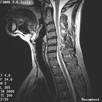
Figure 1: Sagittal T2-weighted fast spin echo (FSE) image in recumbent position
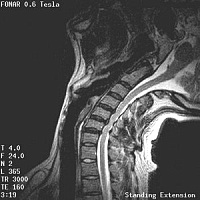
Figure 2: Sagittal T2-weighted FSE image in standing position during extension The gradient recalled echo T2*-weighted axial ima
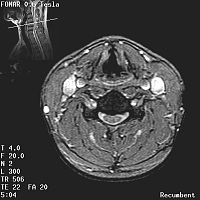
Figure 3: Axial T2*-weighted gradient recalled echo (GRE) image of patient in a recumbent position
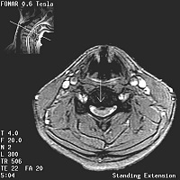
Figure 4: Axial T2*-weighted GRE image of patient in standing-extension
Diagnosis
Fluctuating intervertebral disc herniation dependent upon patient position and dynamic physical maneuver.
Professor J. Randy Jinkins, MD, FACR, FEC
Department of Radiology
Downstate Medical Center
State University of New York
450 Clarkson Avenue
Brooklyn, NY 11203
USA
Clinical Studies performed at:
Melville MRI – Long Island